Medical imaging · 2021–2026
Deep learning for kidney disease imaging
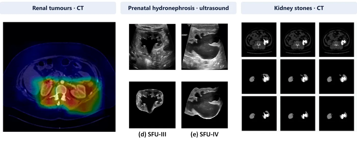
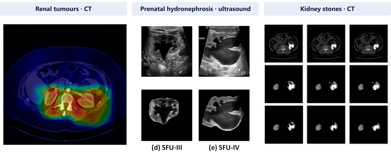
Classifying renal tumours and segmenting kidney stones on CT, and grading prenatal hydronephrosis on ultrasound.
Qatar University MSc research and the HMC–QU–Sidra collaboration, published in three first-author Q1 journal papers
Approach
The project applies deep learning to three separate kidney conditions, each with its own imaging modality, cohort and clinical question: renal tumours on contrast-enhanced CT, prenatal hydronephrosis on ultrasound, and kidney stones on unenhanced CT. The studies share one design idea. A full scan contains far more than the structure of interest, so each pipeline first narrows the image to the kidney, then segments or extracts the relevant region, and only then classifies, grades or measures. The three studies are described below in order of publication. Each was evaluated with patients kept separate between training and testing.
- Narrow the scan to the kidney
- Segment the kidney, tumour or stone
- Classify, grade or measure
Renal tumour subtype and surgical approach from CT
Published · Cancers, 2023
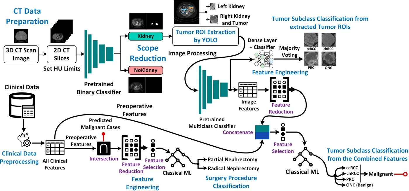
Choosing between partial and radical nephrectomy for a kidney tumour is often unclear. Removing too little risks leaving cancer behind, while removing the whole kidney unnecessarily can lead to lifelong dialysis. The study asked whether combining CT images with clinical data could classify four tumour types and predict the surgical approach. The four types are clear cell, chromophobe and papillary renal cell carcinoma, and oncocytoma.
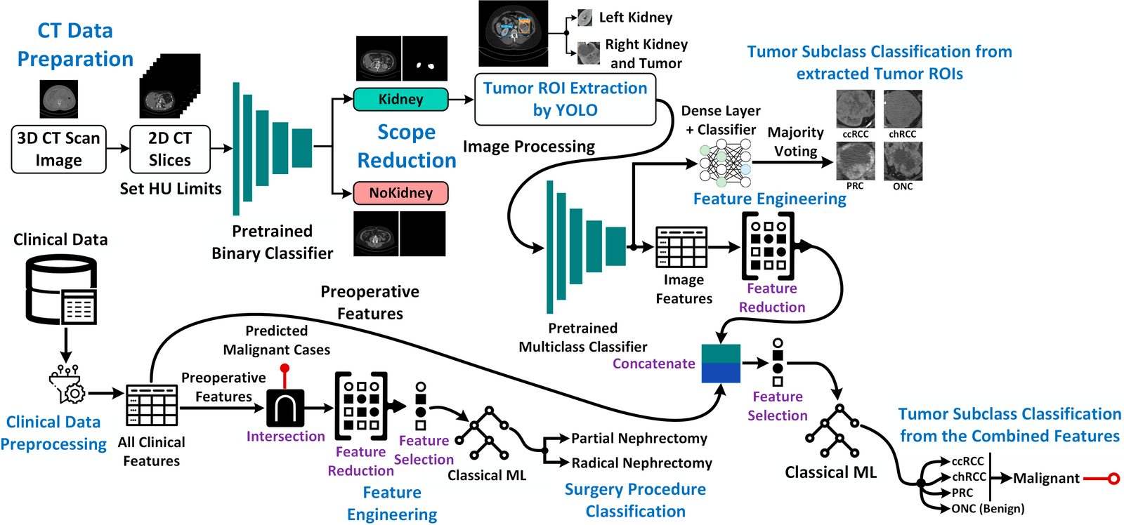
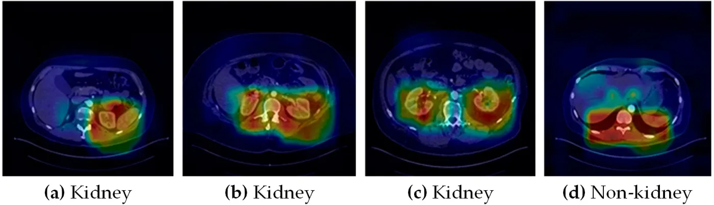
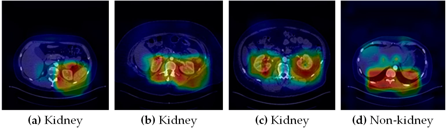
The public KiTS21 dataset provided contrast-enhanced CT and clinical records for 300 patients. Of 64,603 CT slices, about 64.5% contained no kidney, so a modified DenseNet201 classifier with auxiliary outputs, DenseAUXNet201, first removed non-kidney slices. A YOLO detector then extracted tumour regions, and deep image features from those regions were combined with selected clinical variables in the final classifiers. Patients were split into five subject-wise folds.
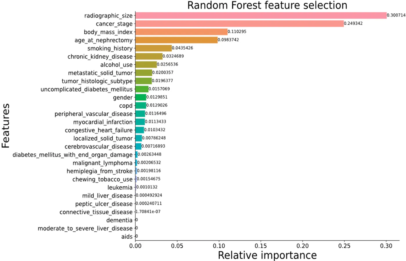
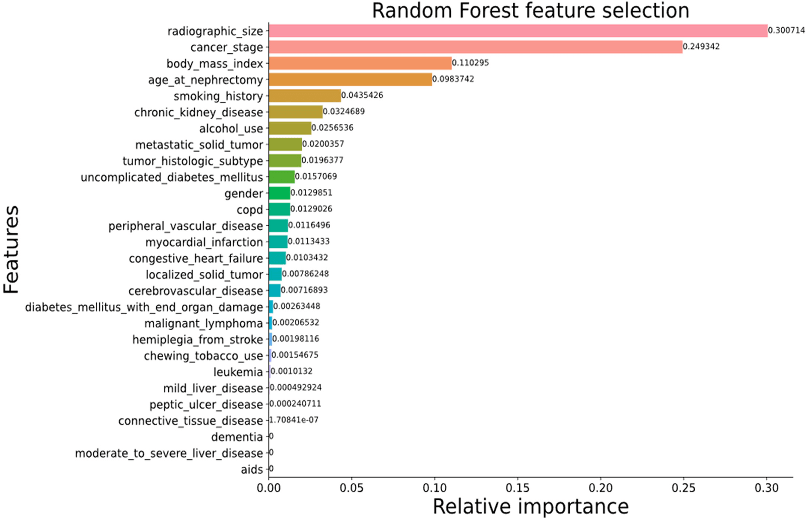
The combined model reached 85.66% accuracy and an 84.92% F1 score for tumour type, despite a marked class imbalance. For malignant tumours, it predicted the recorded surgical approach with 90.63% accuracy. Feature ranking identified tumour size and cancer stage as the most informative clinical variables for the surgical decision.
- Four-class tumour type accuracy
- 85.66%
- Surgical approach prediction accuracy
- 90.63%
- Patients in the KiTS21 cohort
- 300
Grading prenatal hydronephrosis from ultrasound
Published · Expert Systems with Applications, 2024
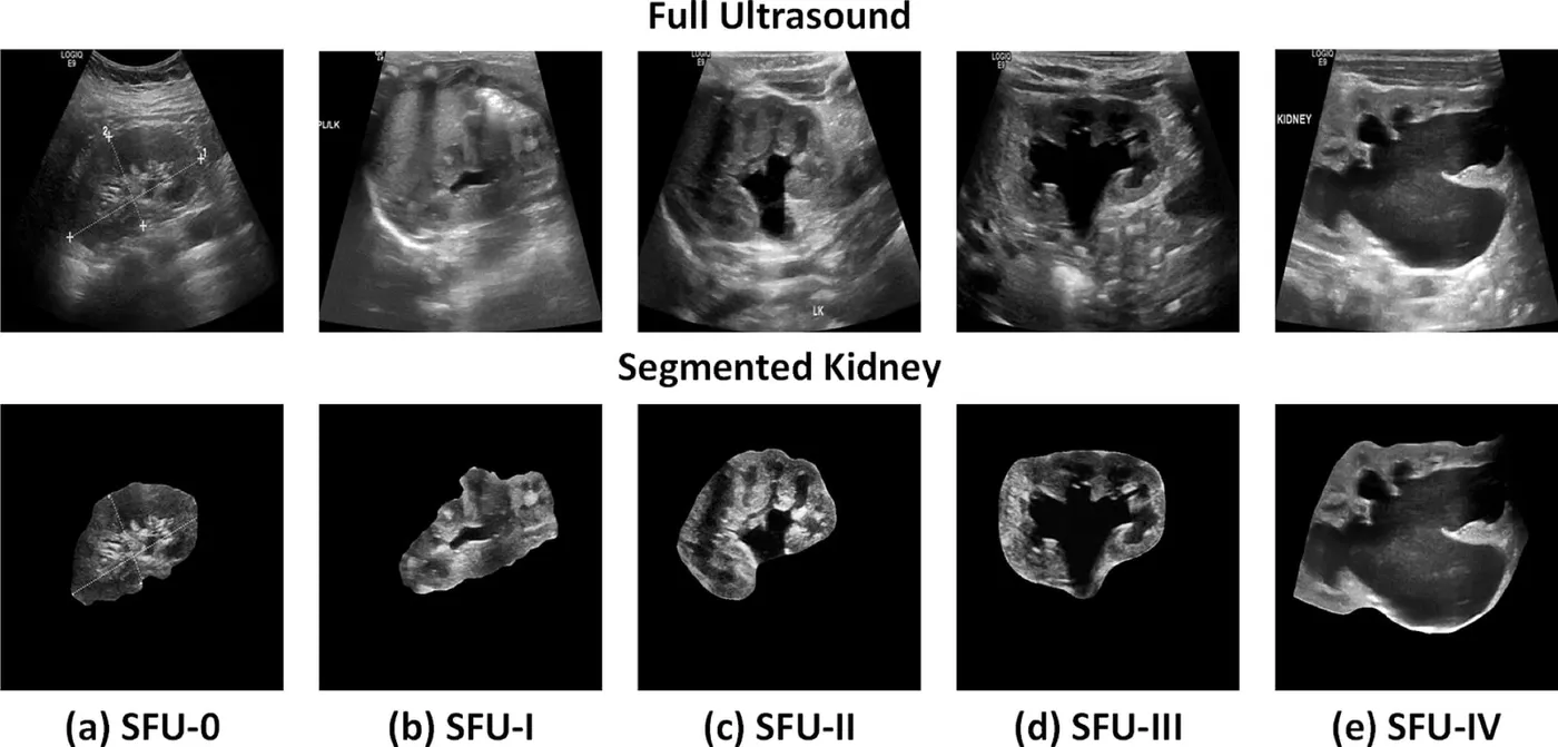
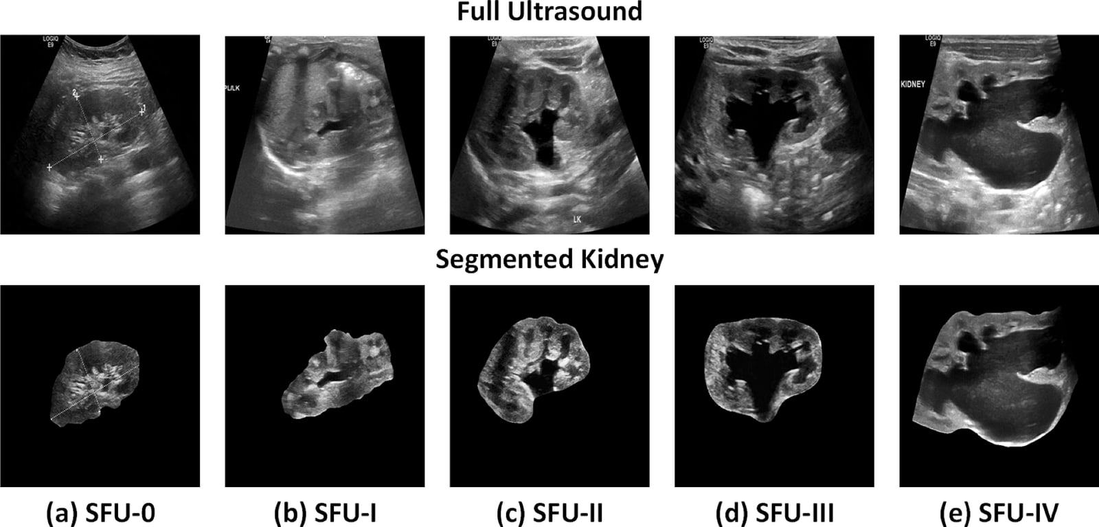
Prenatal hydronephrosis, a swelling of the fetal kidney, is usually benign but can cause lasting damage in severe cases. It is graded from ultrasound on the five-level Society for Fetal Urology (SFU) scale, and that grading varies between clinicians. The study asked whether an automated pipeline could grade severity consistently and support decisions about surgery.
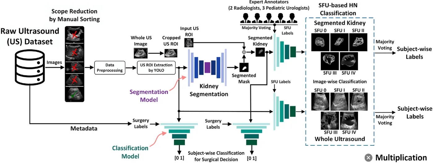
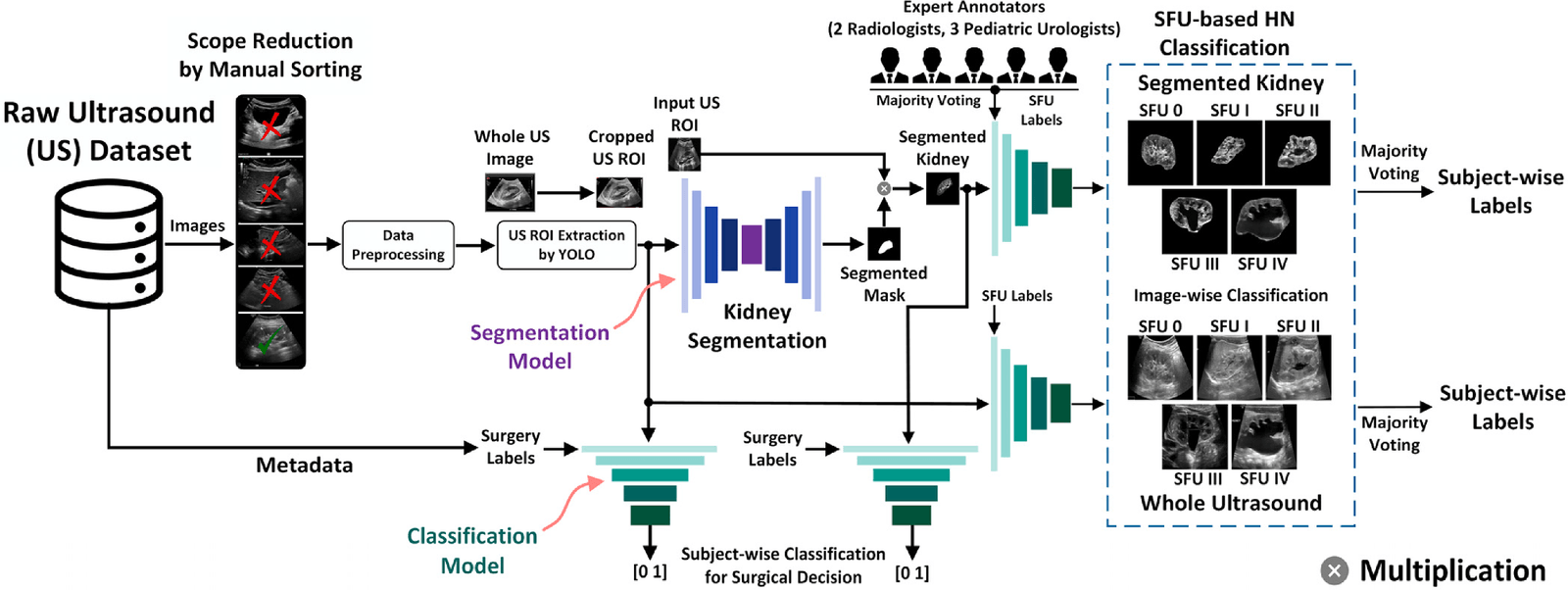
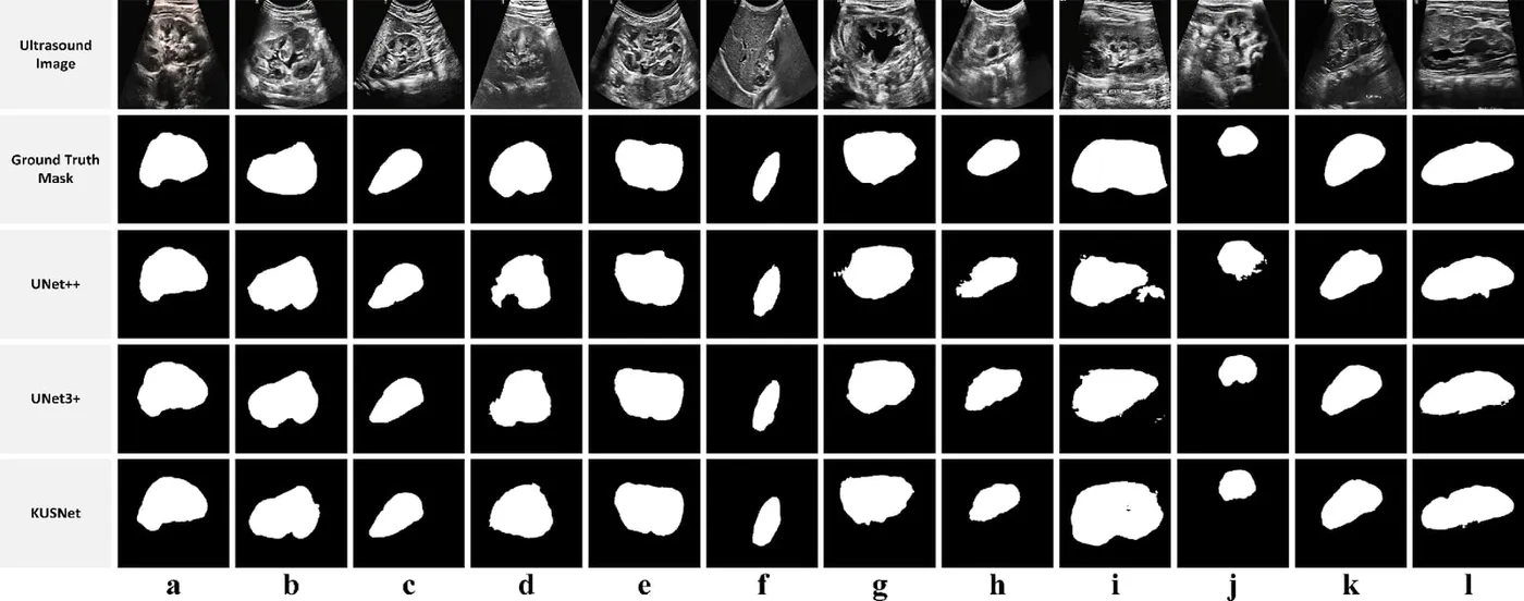
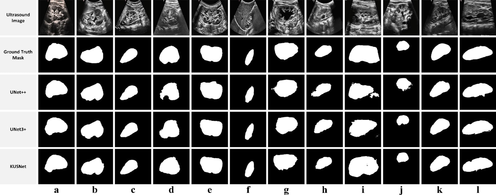
The QU–HMC–AHN dataset was curated with Hamad Medical Corporation, Sidra Medicine and Qatar University from about 5,600 ultrasound images of 164 subjects. After curation, 2,062 images from 163 subjects were used. Two radiologists outlined the kidneys, and two senior radiologists and three senior urologists assigned SFU grades. The pipeline detects the kidney region with YOLOv5x, segments the kidney with the proposed KUSNet, and grades severity with PHCNet, which fuses DenseNet201 and InceptionV3. Weighted voting combines the views of each subject into one grade.
KUSNet segmented the kidney with a 92.1% Dice score. Grading the segmented kidneys reached 93.9% subject-level accuracy and an 89.0% F1 score, and segmenting first improved grading compared with using whole ultrasound images. A separate classifier used the segmented kidneys to separate subjects who went on to surgery from those who did not.
- Kidney segmentation Dice
- 92.1%
- Subject-level five-grade accuracy
- 93.9%
- Subjects in the curated cohort
- 163
Segmenting kidney stones in CT by successive scope reduction
Published · Biomedical Signal Processing and Control, 2026
Kidney stones recur, so patients often need periodic CT monitoring of stone size and position. Stones occupy a tiny fraction of an abdominal CT scan, which makes direct segmentation unreliable. Saliency maps only give a rough location without the stone’s size, shape or exact position. The study asked whether narrowing the search in two steps would let a model outline stones accurately.
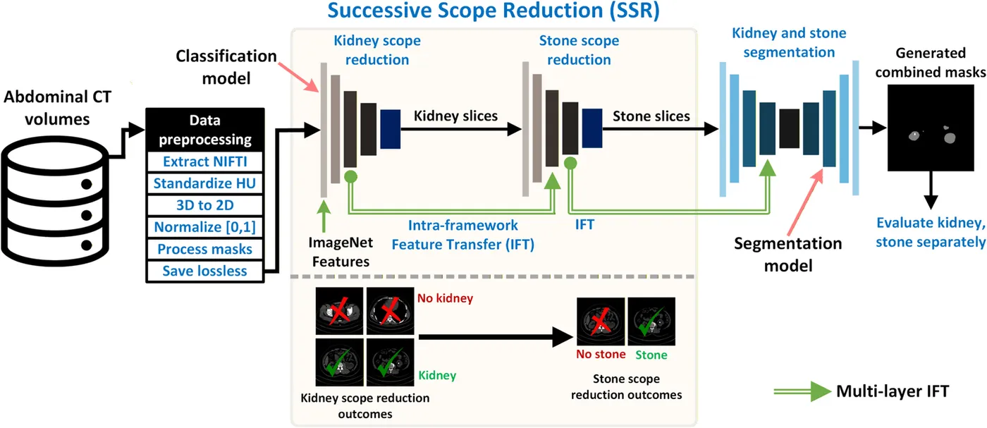
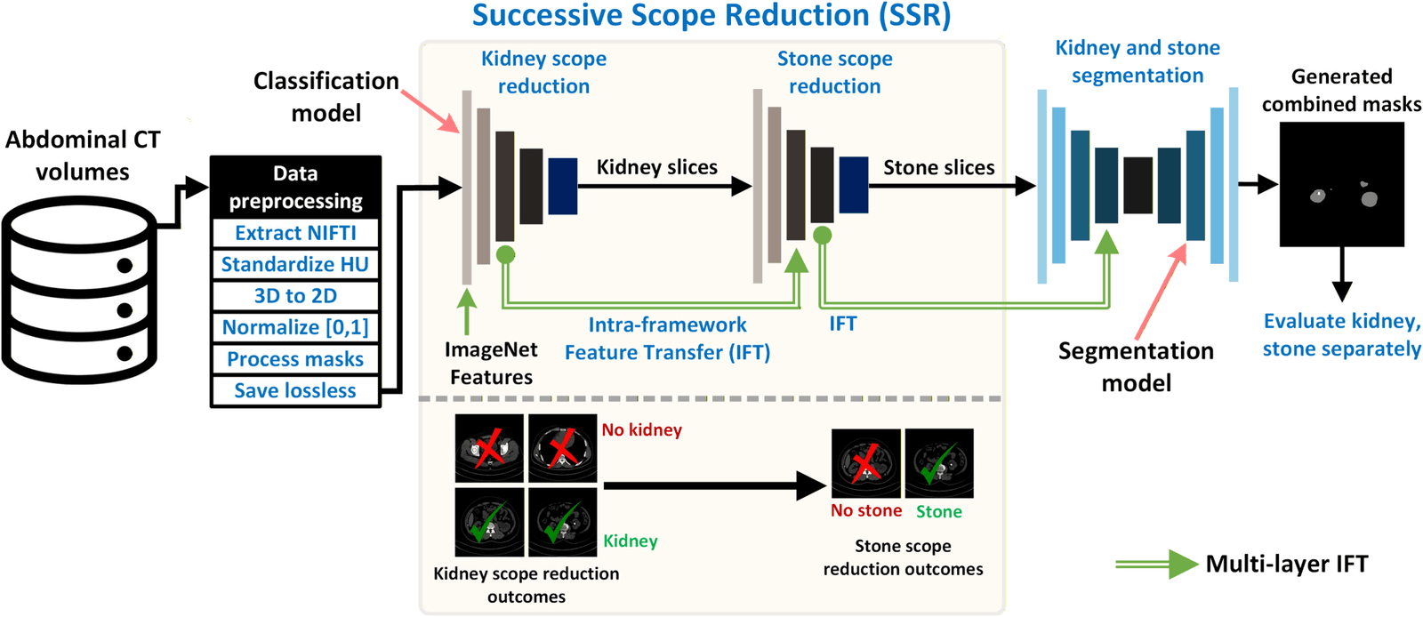
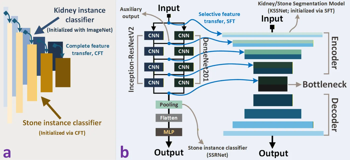
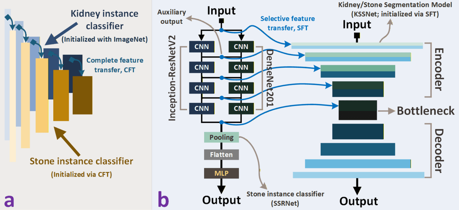
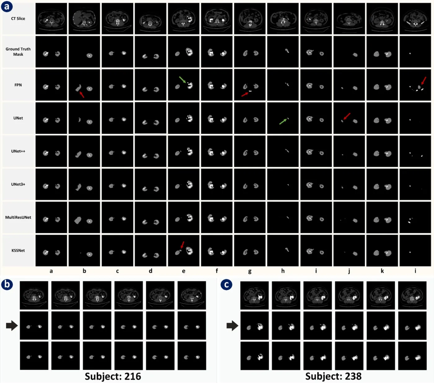
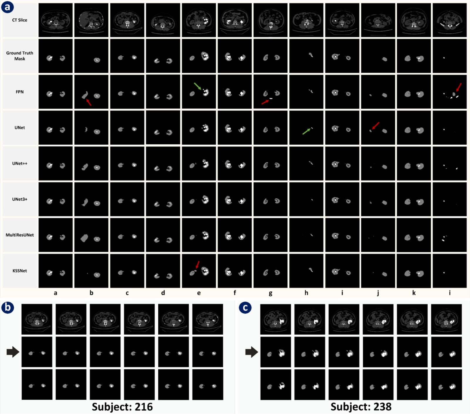
The public dataset contains unenhanced CT from 259 subjects collected in 2018–2020 at the First Affiliated Hospital of Guangzhou Medical University. Successive scope reduction first keeps the 27.55% of the 64,988 slices that contain a kidney, then keeps those with stones. Both steps use SSRNet, which fuses DenseNet201 and Inception-ResNetV2 with auxiliary outputs. Features learned by the classifiers are transferred to initialise KSSNet, a MultiResUNet-based segmentation network. Evaluation used five subject-independent folds.
SSRNet reached a 94.45% F1 score for scope reduction. On the reduced slices, KSSNet segmented stones with a 97.44% Dice score and a 95.01% Jaccard index. The same model reached 90.13% Dice without scope reduction and 93.10% with kidney-slice selection alone. Ablation studies confirmed the contribution of each stage.
- Stone Dice with full scope reduction
- 97.44%
- Stone Dice without scope reduction
- 90.13%
- Scope-reduction F1 score (SSRNet)
- 94.45%
Research stages and outputs
Renal tumour classification and surgical approach modelling
Combined contrast-enhanced CT features and clinical variables from the KiTS cohort to classify renal tumour types and model the surgical approaches recorded in the dataset.
Prenatal hydronephrosis detection and severity grading
Hamad Medical Corporation, Qatar University, and Sidra Medicine contributed to the clinical collaboration. The study used the QU–HMC–AHN ultrasound benchmark and evaluated kidney-region detection, kidney segmentation, and severity grading.
Successive scope reduction for kidney stone segmentation
Narrowed abdominal CT scans to kidney regions and candidate stones, with feature transfer between stages to support segmentation of small targets. The paper appeared after the MSc was completed.
My contribution
All three are first-author papers. The renal tumour study came from my MSc thesis, and I covered its conceptualisation, methodology, software, validation, analysis, data curation, visualisation and writing. The kidney stone study grew from the same thesis research: I designed the successive scope-reduction and feature-transfer pipeline and led model development, experiments, validation, analysis and writing. The hydronephrosis study came from the HMC–QU–Sidra collaboration. As its research assistant I coordinated ethics approval and data acquisition, curated the ultrasound benchmark, developed the detection, segmentation and grading pipeline with KUSNet and PHCNet, and drafted the manuscript.
Scope and validation
The three studies address different clinical questions and use different cohorts and endpoints. The hydronephrosis study was supported by a separate HMC internal grant and also contributed to my MSc thesis.
Next steps
The segment-first approach continues in ongoing work on liver and tumour segmentation across contrast-enhanced CT phases. Further validation of the kidney models on multi-centre cohorts and acquisition protocols would be needed before clinical use.
Publications
- Kidney Cancer Diagnosis and Surgery Selection by Machine Learning from CT Scans Combined with Clinical MetadataCancers · 2023
- Automated grading of prenatal hydronephrosis severity from segmented kidney ultrasounds using deep learningExpert Systems with Applications · 2024
- Enhancing kidney stone segmentation in computed tomography images through successive scope reduction and intra-framework feature transferBiomedical Signal Processing and Control · 2026
Code
- AHN-Severity-StratificationResearch software and repository details
- Kidney-Stone-SegmentationResearch software and repository details