Medical imaging · 2022–2023
Grading prenatal hydronephrosis from ultrasound
Combining multiple fetal kidney ultrasound views into one severity grade, with the kidney outlined before classification.
Hamad Medical Corporation, Sidra Medicine and Qatar University
Approach
Can a pipeline use segmented kidneys across several ultrasound views to estimate a subject-level severity grade?
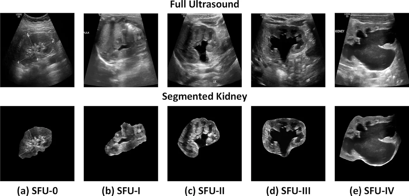
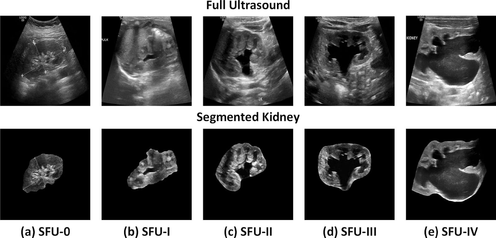
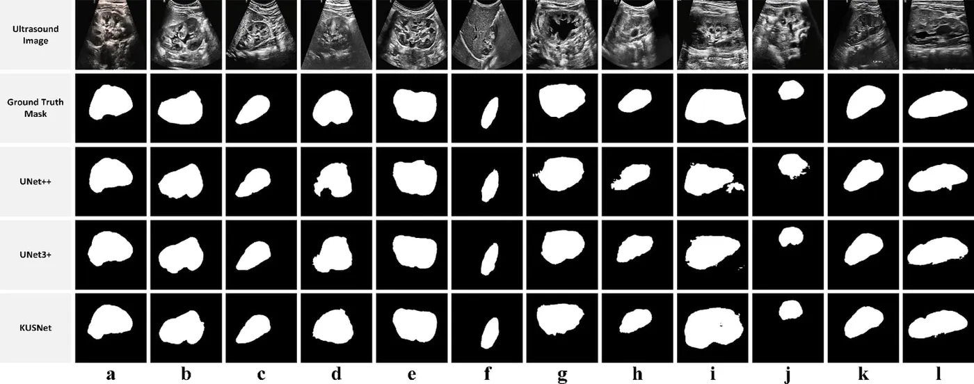
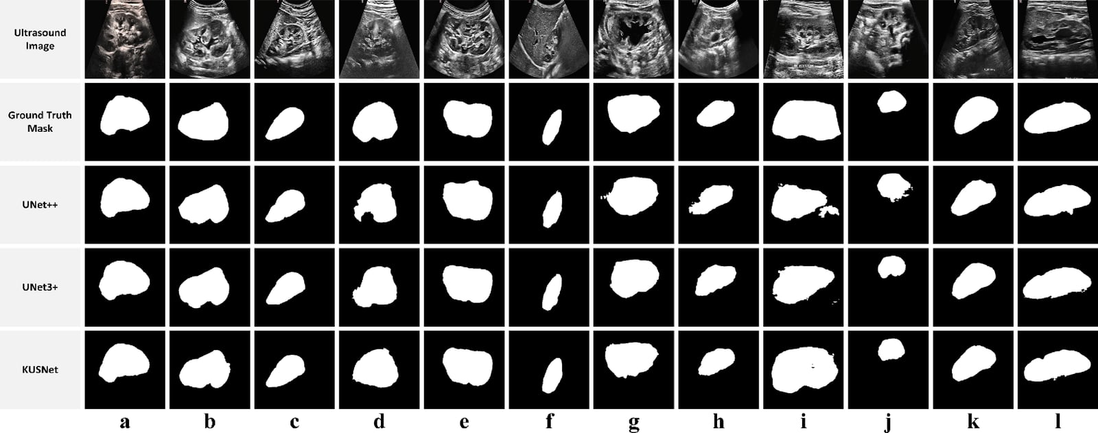
The study collected ultrasound images from 164 subjects. After curation, 2,062 images from 163 subjects were annotated for the kidney region. The workflow locates the region, outlines it with KUSNet, and predicts one of five severity grades with PHCNet. Weighted voting combines grades across ultrasound views at the subject level.
- Locate the kidney region
- Segment with KUSNet and grade with PHCNet
- Combine views into one grade
Illustrated example
From ultrasound views to one severity grade
- Locate the kidney
Find the kidney region in each view.
- Segment and grade each view
KUSNet outlines the kidney. PHCNet grades the segmented views. In this example all three give grade II.
- Combine the grades
Weighted voting gives one subject-level result: Society for Fetal Urology grade II.
Reported results
KUSNet segmented the kidney with a 92.1% Dice score. With segmented views, five-grade classification reached 93.9% subject-level accuracy and an 89.0% F1 score. The two tasks were evaluated separately.
- Kidney segmentation Dice
- 92.1%
- Subject-level grading accuracy
- 93.9%
- Subjects in the curated cohort
- 163
My contribution
I contributed to study design and data curation, developed the methods and software, and wrote the original draft with the research team.
Study scope
Segmentation and grading used five subject-independent folds on the retrospective QU–HMC–AHN cohort. The earlier region-detection stage used a separate two-fold evaluation. Reported grading performance reflects the curated views and reference grades in that cohort.
Publications
- Automated grading of prenatal hydronephrosis severity from segmented kidney ultrasounds using deep learningExpert Systems with Applications · 2024
Code
- AHN-Severity-StratificationResearch software and repository details