Medical imaging · 2026
Kidney stone segmentation in CT
Finding the CT slices most likely to contain kidney stones before tracing their boundaries.
Qatar University MSc research · Qatar University Student Grant QUST-1-CENG-2023-795 and Medical Research Council grant MRC01-21-1027
Approach
Can a model delineate small kidney stones more accurately by narrowing the search before segmentation?
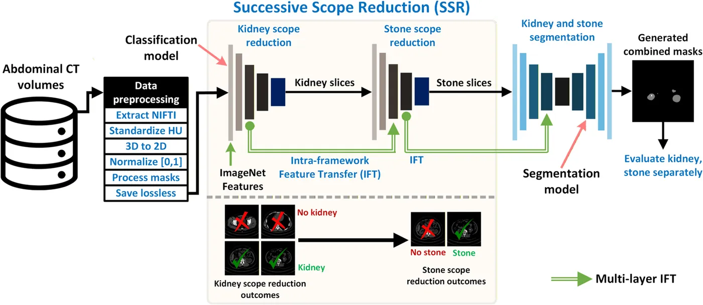
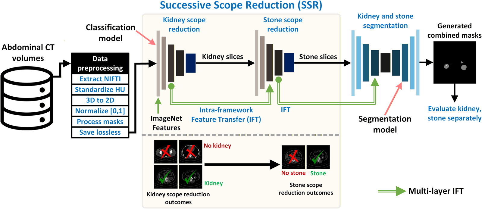
Two classifiers first identify kidney-containing slices and then stone-containing slices. The kidney stone segmentation network, KSSNet, processes the selected images and reuses features learned from kidney tissue segmentation.
- Identify kidney-containing CT slices
- Select slices containing stones
- Trace the stone boundary with KSSNet
Reported results
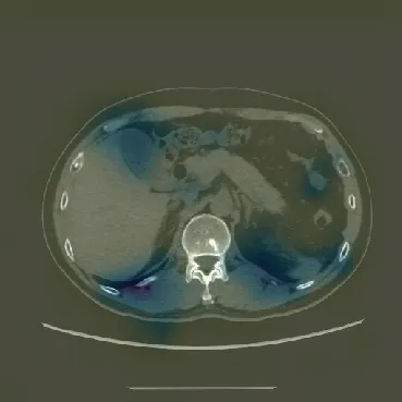
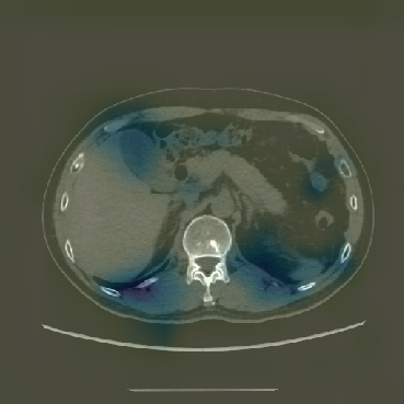
Slice selection improved the same KSSNet model. Its stone Dice score rose from 90.13% without scope reduction to 93.10% with kidney-slice selection only and 97.44% with both stages. The slice classifier reached an overall F1 score of 94.45%.
- Dice with full scope reduction
- 97.44%
- Dice without scope reduction
- 90.13%
- Subjects in the source CT cohort
- 259
My contribution
I designed the successive scope-reduction and feature-transfer approach and led model development, experiments, analysis, and manuscript preparation.
Study scope
The five folds separate subjects. The scans came from a single hospital, so the reported scores describe that cohort and protocol.
Publications
- Enhancing kidney stone segmentation in computed tomography images through successive scope reduction and intra-framework feature transferBiomedical Signal Processing and Control · 2026
Code
- Kidney-Stone-SegmentationResearch software and repository details