Physiological signal processing · 2024
ECG estimation from wearable mechanocardiograms
Reconstructing ECG waveforms from chest-worn accelerometer and gyroscope signals with a deeply supervised 1D segmentation network, then recovering heart rate and its variability.
Qatar University, University of Dhaka and Prince Sattam Bin Abdulaziz University · Prince Sattam Bin Abdulaziz University project PSAU/2023/R/1444
Approach
Can chest vibration and rotation signals from a wearable motion sensor reconstruct an ECG accurate enough to measure heart rate and its variability?
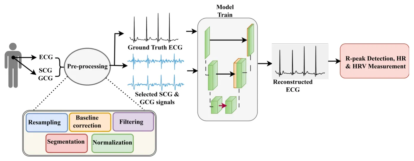
Wearable ECG relies on electrodes that can loosen or degrade during long use. A chest-worn motion sensor can instead record seismocardiograms (SCG), the chest vibrations from each heartbeat, and gyrocardiograms (GCG), the chest rotations; together these are called mechanocardiograms. M2ECG treated translation from mechanocardiogram to ECG as a 1D segmentation problem, so that one motion sensor could provide an ECG-like signal for cardiac monitoring.
- Preprocess SCG and GCG
- Reconstruct ECG with DS-SA-UNet
- Measure heart rate and variability
Choosing mechanocardiogram channels
IEEE Access · 2024
The public Mechanocardiograms with ECG Reference dataset provides lead II ECG with three-axis SCG and GCG from a chest-mounted motion sensor, recorded at 800 Hz. The study used the 29 healthy volunteers, about 260 minutes of recordings.
Signals were band-pass filtered, corrected for baseline drift, resampled and cut into segments of 1,024 samples. Twenty-six volunteers formed the training set, and three held-out volunteers formed the test set.
Of the six motion channels, the SCG z-axis combined with the GCG y-axis gave the lowest reconstruction error, better than all three axes of either sensor or all six channels together.
- Healthy volunteers
- 29
- Volunteers held out for testing
- 3
- Channels used: SCG z-axis and GCG y-axis
- 2
| Volunteers | Segments | |
|---|---|---|
| Training | 26 | 6,214 |
| Test | 3 | 1,503 |
Reconstructing ECG with a deeply supervised SA-UNet
Model comparison
A 1D spatial-attention U-Net with deep supervision (DS-SA-UNet) mapped the two motion channels to ECG. Outputs from deeper decoder levels added weighted losses during training, and the model was trained with the Adam optimiser at a learning rate of 10⁻⁵ and a batch size of four for 100 epochs.
On the held-out volunteers, reconstructed and reference ECG correlated at 81.76% on average, ahead of SA-MultiResUNet at 76.83% and Self-SA-UNet at 71.14%.
- Mean correlation with the reference ECG
- 81.76%
- 1D segmentation models compared
- 3
| Pearson correlation (%) | |
|---|---|
| DS-SA-UNet | 81.76 ± 16.94 |
| SA-MultiResUNet | 76.83 ± 15.83 |
| Self-SA-UNet | 71.14 ± 5.36 |
Recovering heart rate and its variability
Clinical evaluation
R-peaks were detected in the reconstructed and reference ECGs with the Christov algorithm, and mean heart rate and RMSSD were calculated for each of the 1,503 test segments.
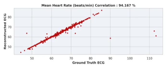
Heart rate from the reconstructed ECG correlated with the reference at 94.17%, compared with 58.57% from SCG alone and 71.92% from GCG alone. For heart rate variability, the correlation rose to 87.07% from 54.75% and 57.53%. For the best test volunteer, the heart rate and variability correlations reached 99.07% and 96.29%.
- Heart rate correlation
- 94.17%
- Heart rate variability correlation
- 87.07%
| SCG | GCG | Reconstructed ECG | |
|---|---|---|---|
| Heart rate | 58.57 | 71.92 | 94.17 |
| Heart rate variability | 54.75 | 57.53 | 87.07 |
My contribution
I contributed to the methodology, data analysis, model evaluation, interpretation and manuscript review.
Study scope
The model was trained and tested on healthy male volunteers at rest, with three volunteers held out in a single split. Recordings from people with coronary artery disease were not used, so reconstruction of abnormal ECGs was not tested. The study presents mechanocardiograms as a complement to ECG rather than a replacement.