Medical imaging · 2023
Cascaded deep learning for liver segmentation from T1-weighted MRI
Benchmarking 24 deep networks and building a cascaded model that handles MRI slices with little visible liver, reaching a 95.15% Dice score.
Qatar University High Impact Grant QUHI-CENG-23/24-216
Approach
Why do liver segmentation networks struggle on some MRI slices, and can a cascade of general and specialised networks fix it?
Outlining the liver on MRI supports the assessment of liver disease, but the organ changes size and shape from slice to slice and sits close to the kidneys and spleen. MRI also has no fixed intensity scale like CT. This study compared a wide range of segmentation networks on T1-weighted scans, identified where the best of them failed, designed a cascade around that weakness, and deployed the model in a cloud prototype.
- Prepare T1-weighted slices
- Benchmark 24 networks
- Cascade general and specialised models
Benchmarking 24 segmentation networks
Five-fold cross-validation on CHAOS
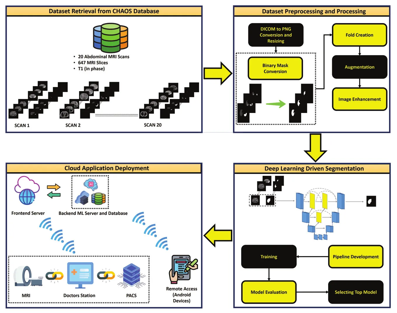
The study used T1-weighted in-phase MRI from the public CHAOS challenge: 647 slices from 20 people, with liver masks annotated by certified radiologists. In-phase T1 images were chosen because fat appears brighter, which gives the networks more contrast to work with. MRI also lacks the fixed intensity scale that makes CT easier to preprocess.
UNet, UNet++ and Feature Pyramid Networks were each paired with eight dense, residual and inception encoders, giving 24 networks. Each was tested on the original slices and on three-channel and gamma-corrected versions. UNet++ with a DenseNet201 encoder performed best on the original images. The enhancement methods slightly lowered performance overall, because they did not help slices where the liver was small.
- Network and encoder combinations compared
- 24
- T1-weighted slices from 20 people
- 647
- Best single-network Dice (UNet++ with DenseNet201)
- 94.30%
A cascade for slices with little liver
Main result
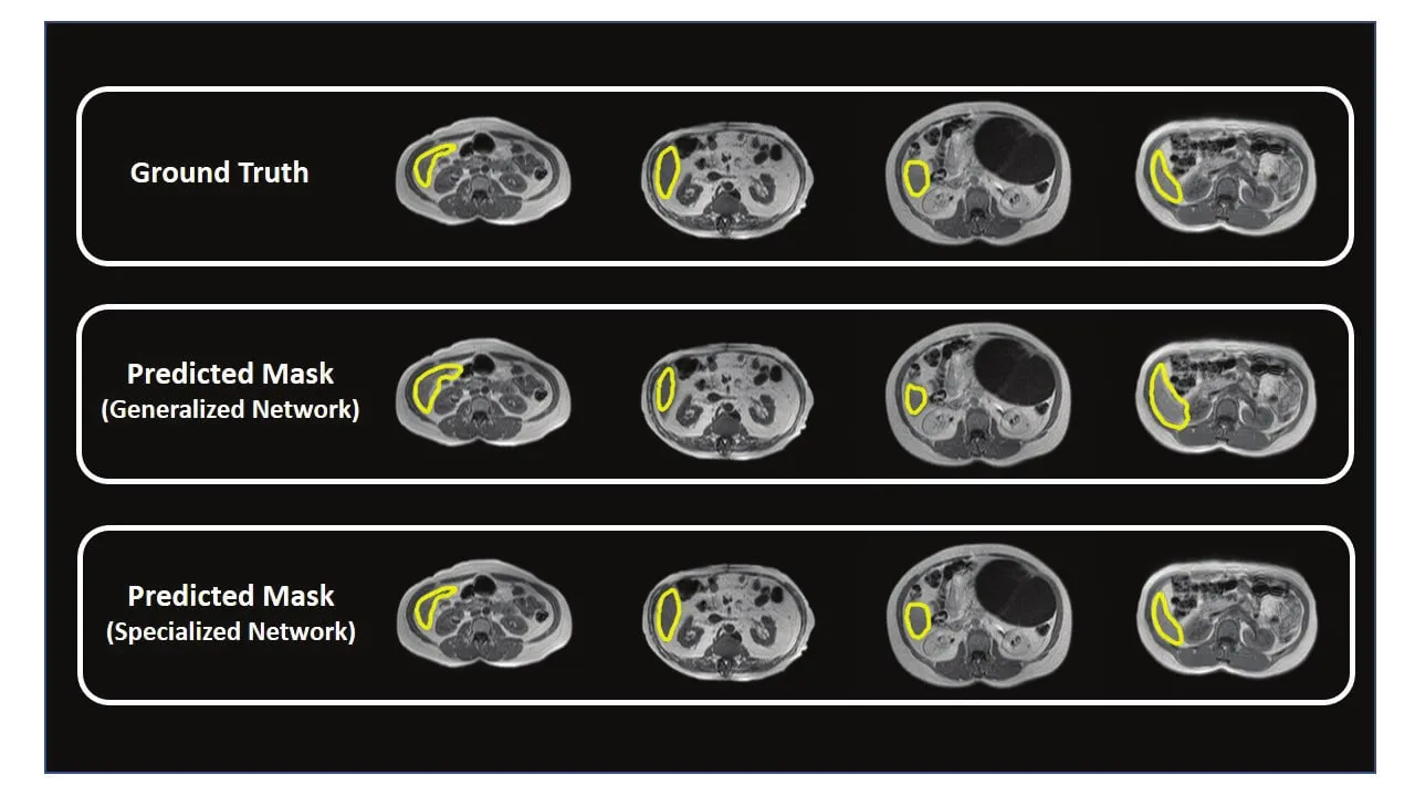
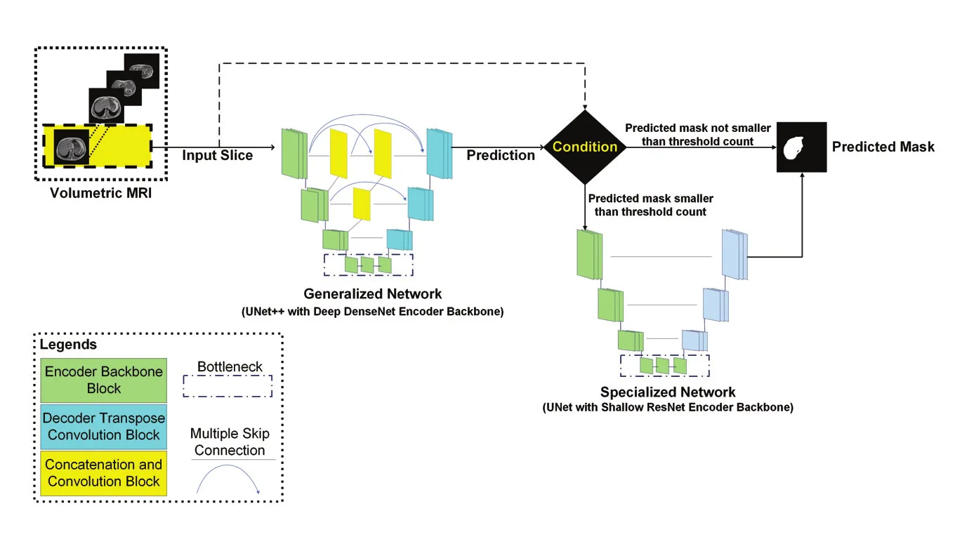
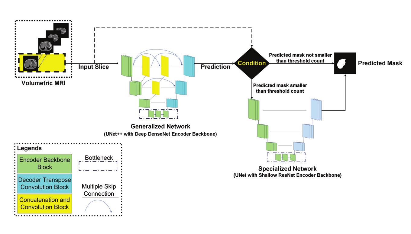
The best single network scored above 93% on slices through the middle and upper liver, but only 78–83% across folds on slices through its lower part, where little liver is visible. The cascade addresses this directly. A generalised network segments every slice. If it finds no liver the slice is left empty, if it finds more than 750 liver pixels its mask is kept, and otherwise a specialised network trained on small-liver slices produces the final mask.
A shallow UNet with a ResNet18 encoder was the strongest specialised network. The full cascade reached a Dice score of 95.15%, higher than every earlier result the paper compared on the same test set, the closest being 93.60% with nnU-Net.
- Liver Dice with the cascade
- 95.15%
- Liver IoU with the cascade
- 92.10%
| Dice | IoU | |
|---|---|---|
| Small-liver slices, best generalised network | 80.88% | 70.74% |
| Small-liver slices, specialised UNet with ResNet18 | 86.22% | 77.00% |
| All slices, UNet++ with DenseNet201 | 94.30% | 91.00% |
| All slices, cascaded network | 95.15% | 92.10% |
A cloud application prototype
Demonstration software
The model was deployed on a Google Cloud server with a database for the images, a browser interface and an Android application. A scheduler was built to retrain the deployed model as users add data, and the design allows a later connection to a hospital picture archiving system.
My contribution
I contributed to the software implementation and data curation, helped draft the original manuscript, and took part in reviewing and editing it.
Study scope
The study used one public dataset of 20 people without liver disease and evaluated 2D slices with cross-validation, so performance on liver lesions, other scanners and independent cohorts remains to be tested. Multiphase liver and tumour segmentation on CT is the focus of LiPIVOT.