Physiological signal processing · 2023–2024
Deep learning for non-invasive fetal ECG extraction
Deep learning methods that recover fetal ECG waveforms and heartbeat timing from abdominal recordings containing maternal ECG and noise.
Qatar National Research Fund NPRP 10-0123-170222 · Qatar University QUHI-CENG-22/23-548
Approach
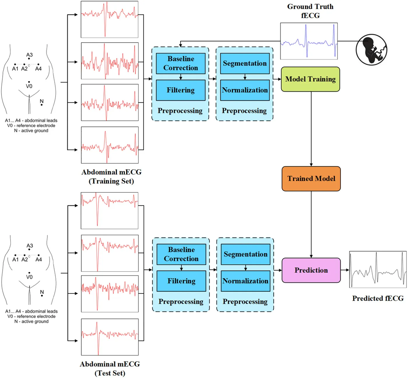
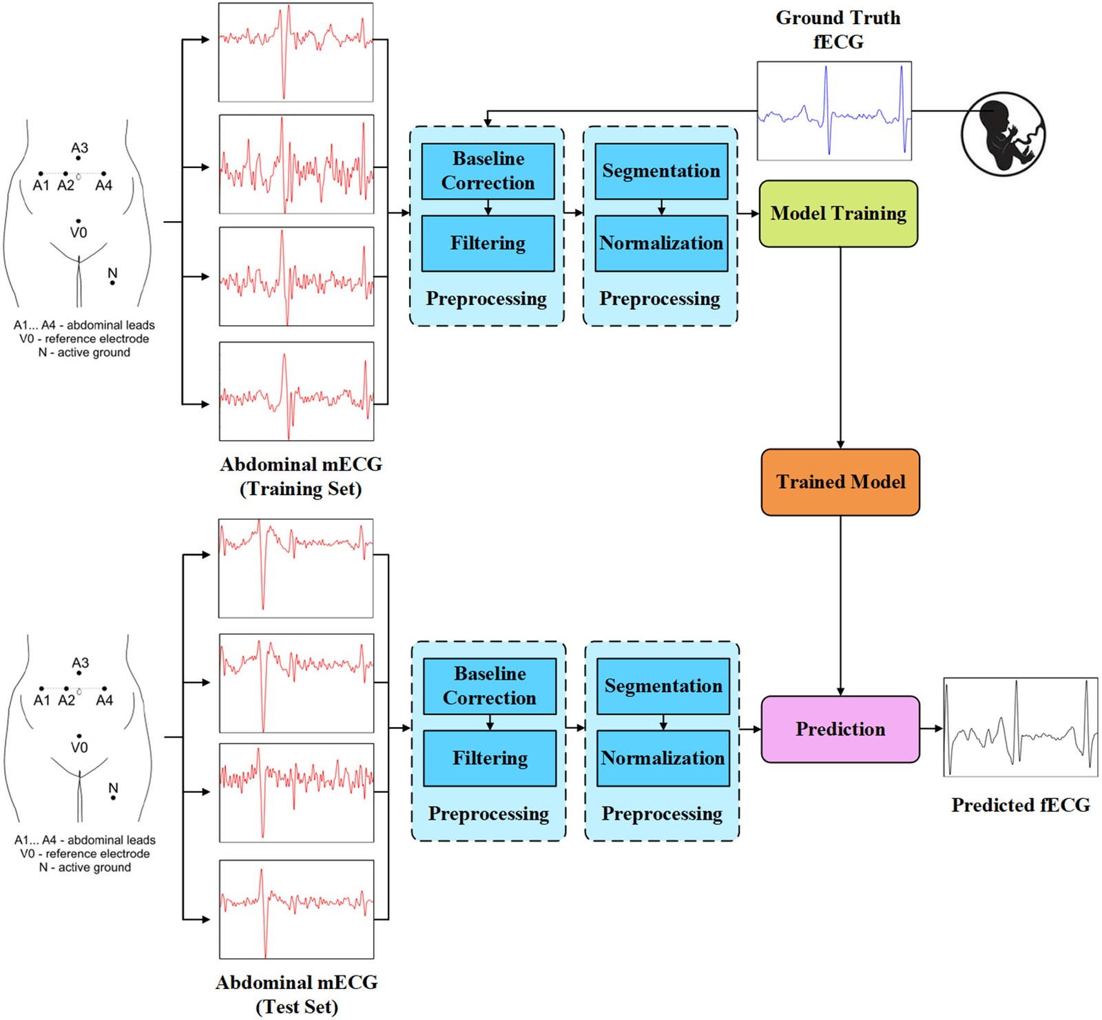
Can fetal ECG morphology and heartbeat timing be recovered from electrodes placed on the mother’s abdomen?
Fetal scalp electrodes provide a clear ECG, but they are invasive and can only be used around delivery. Abdominal electrodes are non-invasive, although the weak fetal signal overlaps with the stronger maternal ECG and noise. This project first used a deeply supervised segmentation network to extract fetal ECG, then used a 1D-CycleGAN to better preserve waveform morphology and estimate fetal heart measures.
- Record abdominal maternal ECG
- Reconstruct fetal ECG
- Evaluate waveforms and heartbeats
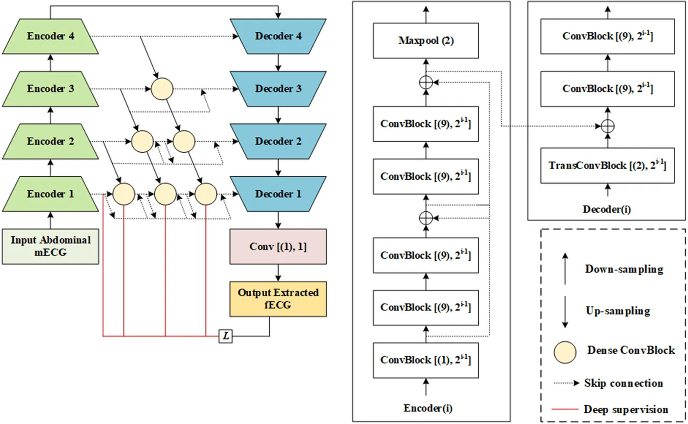
Extracting fetal ECG with deeply supervised LinkNet++
Engineering Applications of Artificial Intelligence · 2023
The study used two public datasets of four-channel abdominal ECG. Dataset 1 contains recordings from five women at 38 to 41 weeks of pregnancy, and Dataset 2a contains recordings from 12 women during labour, both with fetal scalp ECG as the reference. Dataset 2b adds recordings from 10 women with expert-marked fetal heartbeats.
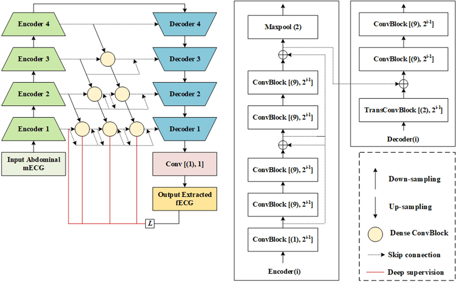
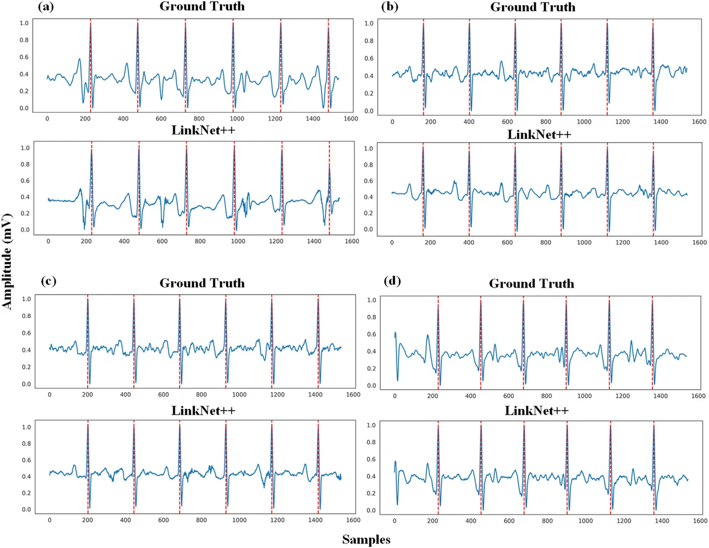
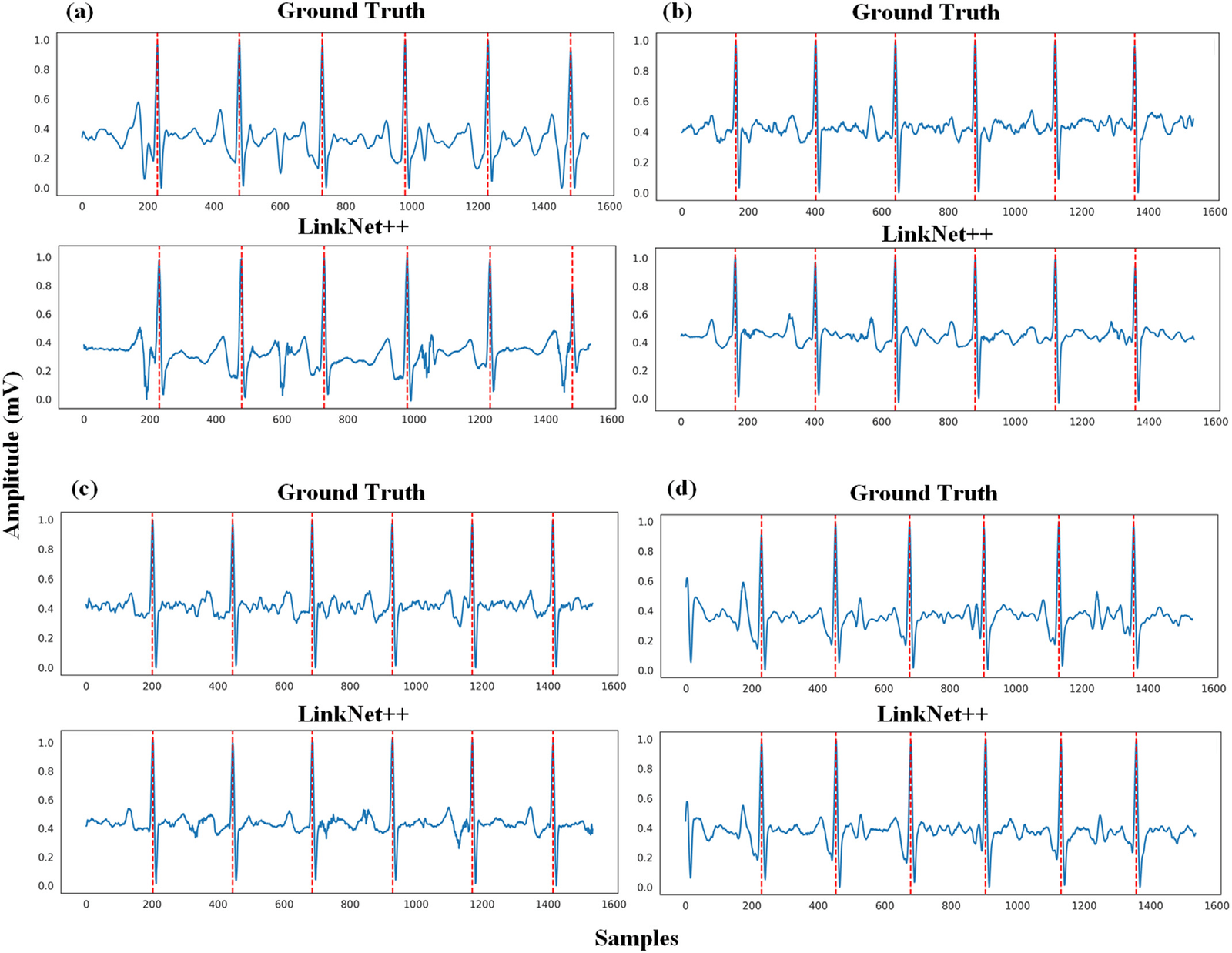
LinkNet++ combines LinkNet’s residual encoder and feature addition with dense skip pathways and deep supervision. It was compared with U-Net, UNet variants and LinkNet using leave-one-subject-out validation. LinkNet++ gave the highest waveform correlation, improving on LinkNet by 3.56 points on Dataset 1 and 2.58 points on Dataset 2a.
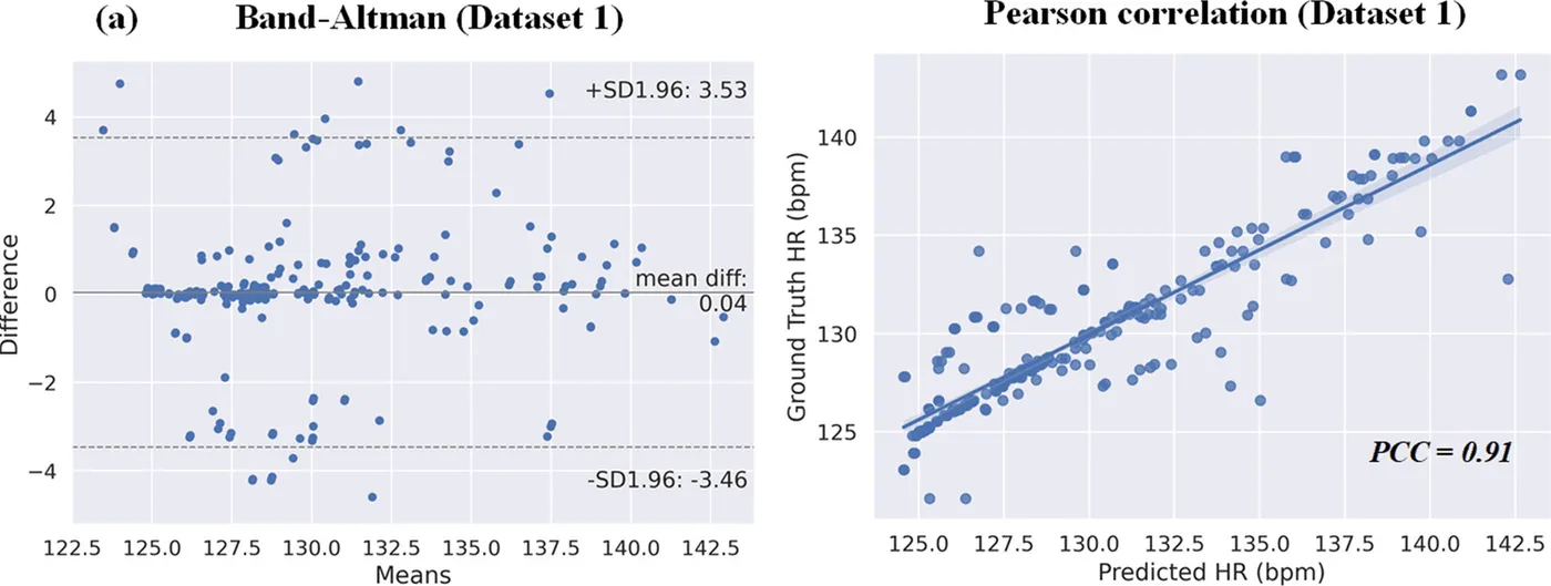
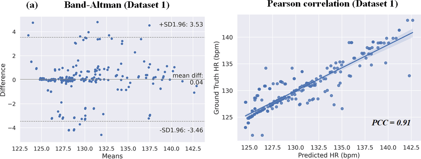
Fetal QRS complexes detected from the extracted signal reached F1 scores above 99% on all three datasets. Heart rate from the extracted signal also correlated with the reference at more than 0.9.
- Best waveform correlation (Dataset 2a)
- 87.60%
- Best fetal QRS F1 score (Dataset 1)
- 99.6%
- Women across the three datasets
- 27
| Waveform correlation | Mean absolute error | QRS precision | QRS sensitivity | QRS F1 | |
|---|---|---|---|---|---|
| Dataset 1 | 85.58% | 0.068 | 99.7% | 99.5% | 99.6% |
| Dataset 2a | 87.60% | 0.061 | 99.5% | 99.3% | 99.4% |
| Dataset 2b | Not available | Not available | 99.4% | 99.6% | 99.5% |
Preserving fetal ECG morphology with 1D-CycleGAN
Expert Systems with Applications · 2024
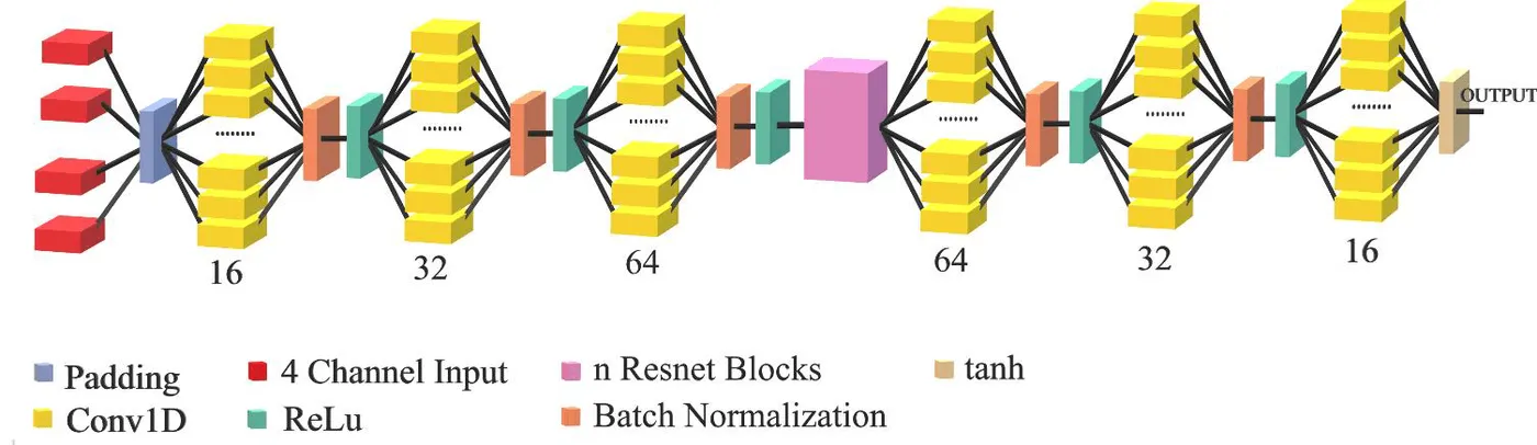
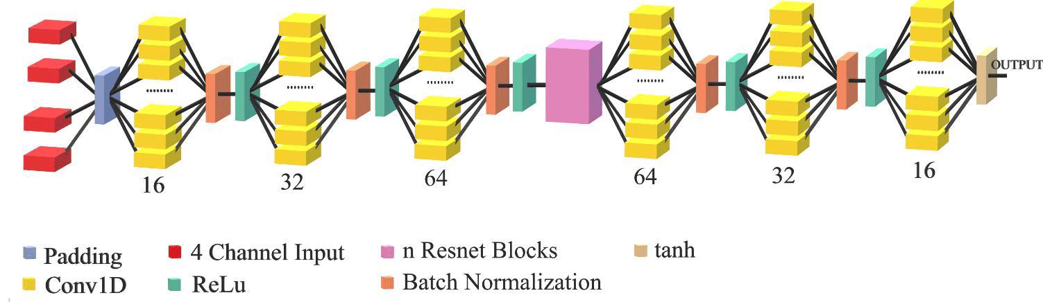
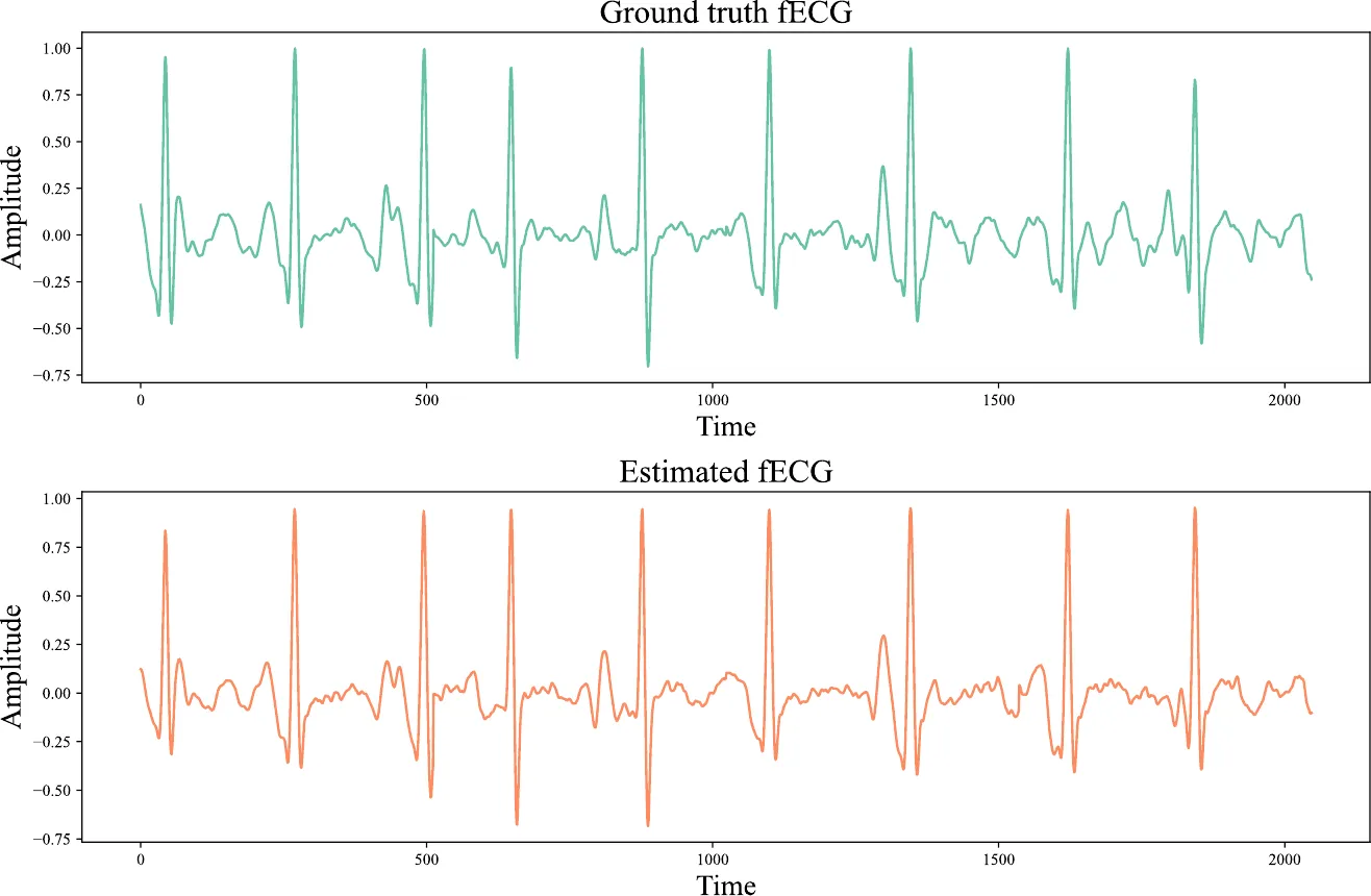
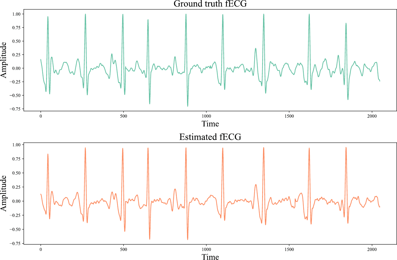
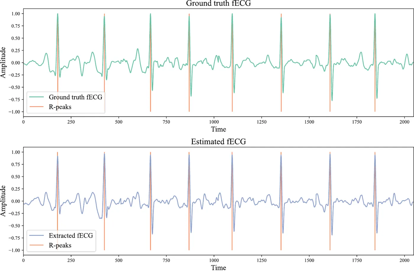
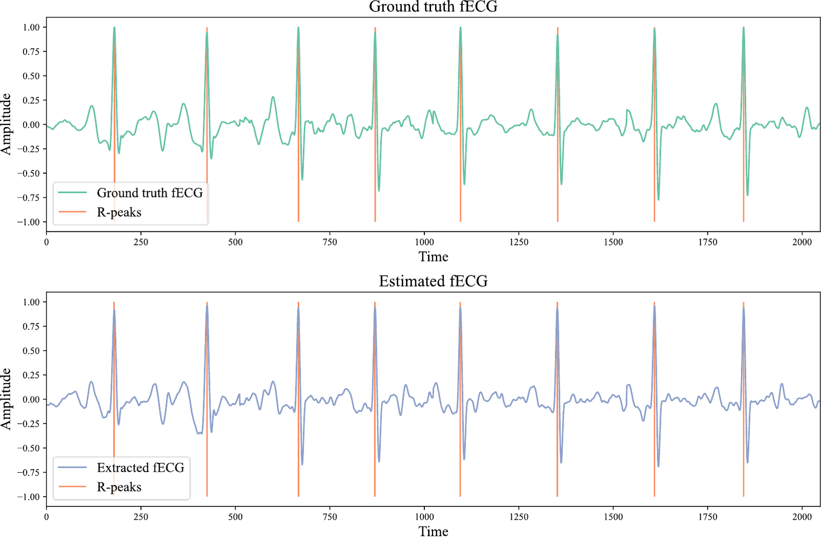
The second study focused on the full waveform, including features such as P waves, ST segments and T waves. A 1D-CycleGAN learned to map four-channel maternal ECG to fetal ECG using the two PhysioNet datasets with scalp reference signals. Preprocessing included zero-lag filtering to avoid shifting the waveform.
A weighted loss added spectral, temporal and power terms to the adversarial loss, increasing waveform correlation by almost 10%. Among U-Net, ResNet and Self-FPN generators, a 13-block ResNet generator with a standard PatchGAN discriminator gave the best result. The full network has 0.762 million parameters.
- Average waveform correlation
- 88.4%
- Average spectral correlation
- 89.4%
- ResNet blocks in the best generator
- 13
| Waveform correlation | Spectral correlation | RMSE | Mean absolute error | |
|---|---|---|---|---|
| Fold 1 | 0.89 | 0.90 | 0.106 | 0.069 |
| Fold 2 | 0.88 | 0.88 | 0.109 | 0.073 |
| Fold 3 | 0.89 | 0.90 | 0.103 | 0.068 |
| Fold 4 | 0.89 | 0.90 | 0.102 | 0.069 |
| Fold 5 | 0.87 | 0.89 | 0.115 | 0.077 |
| Mean | 0.884 | 0.894 | 0.107 | 0.071 |
Measuring fetal heartbeats from the extracted signal
Expert Systems with Applications · 2024
Fetal QRS complexes were detected from the 1D-CycleGAN output with a tolerance of 31.25 ms. Mean heart rate and beat interval closely matched the reference, showing that the extracted signal preserved fetal timing. Short-term variation in heart rate was harder to reproduce.
- Fetal QRS F1 score
- 96.4%
- Fetal QRS precision
- 97.6%
- Mean heart-rate error
- 0.25%
| Reference | Extracted | Error | |
|---|---|---|---|
| Mean R-R interval | 473.19 ms | 474.46 ms | 0.27% |
| Mean heart rate | 128.52 bpm | 128.85 bpm | 0.25% |
| Heart-rate standard deviation | 14.14 bpm | 18.24 bpm | 29% |
| SDNN/RMSSD | 0.70 | 0.70 | 0.57% |
| pNN50 | 40.45% | 41.39% | 2.32% |
My contribution
For LinkNet++, I contributed substantially to the methodology, model development, experiments, analysis and validation, and manuscript writing and revision. For the 1D-CycleGAN study, I contributed to the deep learning methodology, model assessment, result analysis and interpretation, and manuscript review.
Study scope
Both studies used small public datasets from late pregnancy and labour, with limited variation in recording conditions. Scalp ECG and expert annotations provided the references. Validation across more participants, earlier gestation, different devices and fetal heart conditions is needed before clinical use.
Publications
- A novel deep learning technique for morphology preserved fetal ECG extraction from mother ECG using 1D-CycleGANExpert Systems with Applications · 2024
- Fetal ECG extraction from maternal ECG using deeply supervised LinkNet++ modelEngineering Applications of Artificial Intelligence · 2023